A Handbook Of Some Common South Indian Grasses: 5-Histology Of The Vegetative Organs
This is an extract from |
Contents |
Histology Of The Vegetative Organs
The shoots and roots of grasses conform in their internal structure to the monocotyledonous type. In all grasses numerous threads are found running longitudinally within the stem and some of these pass into the leaves, at the nodes, and run as nerves in the blades of the leaves. These threads are the vascular bundles. The rest of the tissue of the stem and leaves consists of thin-walled parenchymatous cells of different sorts.
The general structure of these bundles is more or less the same in all grasses. A vascular bundle consists of only xylem and phloëm, without the cambium, and so no secondary thickening can take place in the stems of grasses. Such bundles as these are called closed vascular bundles to distinguish them from the dicotyledonous type of vascular bundles which are called open vascular bundles on account of the existence of the cambium.
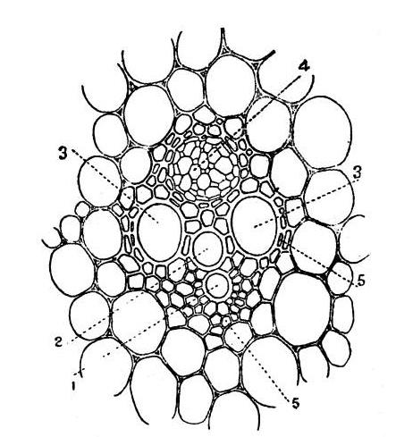
The component parts and elements of which the vascular bundles in grasses are composed may be learnt by studying the transverse and longitudinal sections of these bundles in any grass. The cross and longitudinal sections of a vascular bundle of the stem of Pennisetum cenchroides, are shown in figs. 22 and 23. In the figure of the transverse section the two large cavities indicated by the number 3 and the two small circular cavities with thick walls lying between the larger ones and indicated by the numbers 1 and 2 are the chief elements of the xylem.
By looking at the longitudinal section it is obvious that these elements are really vessels, the larger being pitted and the smaller annular and spiral vessels. These vessels together with the [Pg 20]numerous small thick-walled cells lying between the pitted vessels constitute the xylem. Just above the xylem there is a group of large and small thin-walled cells. This is the phloëm and it consists of sieve tubes and thin-walled cells.
All round the xylem and the phloëm there are many thick-walled cells. These are really fibres forming the bundle-sheath. On account of this bundle-sheath the bundles are called fibro-vascular bundles.
Structure of the stem
Structure of the stem.—The stem of a grass consists of a mass of parenchymatous cells with a number of fibro-vascular bundles imbedded in it, and it is covered externally by a protective layer of cells, the epidermis. The stem is usually solid in all grasses in the young stage, but as it matures the internodes become hollow in many grasses and they remain solid in a few. In the internodes the fibro-vascular bundles run longitudinally and are parallel, but in the nodes they run in all directions and form a net work from which emerge a few bundles to enter the leaves.
So far as the broad general features are concerned, the stems of many grasses are more or less similar in structure. However, when we take into consideration the arrangement of bundles, the development and arrangement of sclerenchyma, every species of grass has its own special characteristics. And these are so striking and constant that it may be possible to identify the species from these characters alone.
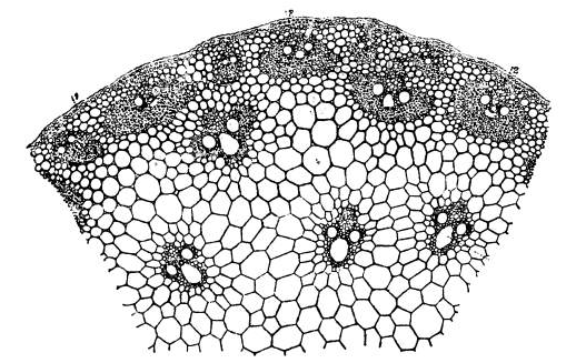
We may take as a type the stem of Rottboellia exaltata. This stem is somewhat semi-circular in transverse section and it is almost straight and flat in the front (the side towards the axillary bud). The peripheral portion of the stem becomes somewhat rigid and thick due to the aggregation of vascular bundles, some small and others large.
The outermost series of bundles consisting of small and larger bundles are in contact with the layers of the cells lying just beneath the epidermis and these cells are also thick-walled. A few are away from these being separated by three or four layers of cells from the peripheral bundles. In all these vascular bundles the bundle-sheath is very strongly developed all round and is very much developed especially at the sides.
It is this great development of sclerenchyma that makes the outer portion of the cortex hard. Within the ground tissue are found a number of vascular bundles scattered more or less uniformly. These bundles have no continuous bundle-sheaths but have instead groups of fibres at the sides and in front of the phloëm. The cavities near the annular vessels are somewhat larger and conspicuous in these bundles.
In order to give a general idea of the variations in the structure of the stem in grasses a few examples are chosen and the details of the structure of the stems of these grasses are dealt with here.
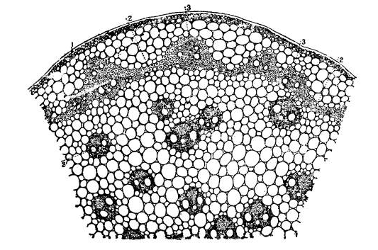
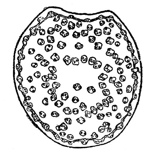
The stem of Pennisetum cenchroides is somewhat round in outline in the transverse section with a slight curvature in the front. The vascular bundles are rather numerous and irregularly scattered all over the ground tissue. The peripheral bundles are not so close to the periphery of the stem as in Rottboellia exaltata. These are separated from the epidermis by several layers of parenchymatous cells. Further, these peripheral bundles are all imbedded in a continuous sclerenchymatous band which runs round the stem in the form of a ring. The epidermal cells as well as the layer of cells in immediate contact with it are thick-walled. In the vascular bundles of the ground tissue the bundle-sheath is rather prominent and the phloëm portion is well developed.
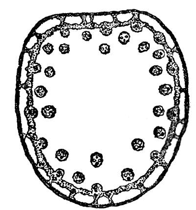
In the stem of Eriochloa polystachya, all the vascular bundles are more or less peripheral in position leaving a wide area of parenchymatous cells in the centre. The outline of the stem in cross section is rotund or ovate-rotund with the front side somewhat flattened and straight. The epidermal cells alone are [Pg 23]thickened. A well developed continuous ring of sclerenchyma is present and this is connected with the epidermal layer at short intervals by means of short sclerenchymatous bands. So the parenchymatous cells of the cortex lying outside the sclerenchymatous ring are divided into small isolated areas. There are three series of vascular bundles.
One series consists of small bundles lying inside the sclerenchyma ring at the base of each of the connecting bands. The second series is made up of large vascular bundles imbedded in the ring so as to bulge out inside the ring. The vascular bundles of the third series are found just away from the ring and separated from it by a few layers of parenchymatous cells.
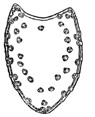
Another stem in which the vascular bundles are more or less peripheral in position and enclosing a wide parenchyma is that of Setaria glauca. In the transverse section of the stem the outline is ovate, laterally compressed, obtusely keeled at the back and somewhat concave in the front. The sclerenchymatous band is narrow and continuous and very close to the epidermis, being separated from it only by two or three layers of thin-walled cells. The epidermal cells alone are thickened. As to the vascular bundles there are three sets. One set of bundles lying just outside the sclerenchymatous ring consists of small ones connecting the ring with the epidermis.
Just inside the sclerenchymatous ring lies a series of bundles which are connected with it. Still inside, at some distance from the sclerenchymatous band, are seen vascular bundles forming a row and enclosing a large space of the ground tissue consisting of only parenchyma. (See figs. 29 and 30.)
The stem of Panicum ramosum is semi-circular and somewhat flat on one side. The epidermal cells alone are thickened. There [Pg 26]is a broad well developed continuous band of sclerenchyma, which is connected at regular intervals with the epidermis by small vascular bundles. Another row of vascular bundles lies just inside the sclerenchymatous ring and each of these bundles is in contact with the band. Away from the ring lie a number of bundles forming a series disposed in two irregular rings around a broad portion of the ground tissue.
The stem of the grass Andropogon caricosus is oval in outline, the front being flat. The epidermal cells and those below and in contact with them are thick-walled. The sclerenchymatous ring though present is very narrow and not very conspicuous. It consists of one or two layers of cells connecting a few vascular bundles forming the outermost set. There is a series of vascular bundles inside the ring which surrounds a large area of the ground tissue. Two isolated bundles, one in front and another at the back of the ground tissue, are found. The cells of the ground tissue lying just inside the vascular bundles are all very much thickened.
The stems of Panicum Isachne and Eragrostis interrupta are hollow. The stem of the former is circular in outline in cross section, though wavy. There is a sclerenchymatous ring close to the epidermis but separated from it by a few layers of parenchyma. One set of bundles is imbedded in the band, and another set just touches the inner border of it. A third series is disposed around a fairly large amount of ground tissue, which may or may not have a cavity in the centre.
The stem of Eragrostis interrupta has more or less the same structure, but the cortex has air spaces here and there. Other minor differences may be seen on referring to figs. 35 and 36.
The stems of grasses growing in wet or marshy situations differ in structure from those detailed above. As examples the stems of Panicum flavidum, Panicum colonum, Panicum Crus-galli and Panicum fluitans may be considered. The stem of Panicum flavidum is broadly ovate in cross section with a flat front and is more or less solid, though occasionally the parenchymatous cells in the centre get broken.
Two rows of vascular bundles surround a fairly large amount of parenchymatous cells of the ground tissue. There is a continuous ring of sclerenchyma separated from the epidermis by a fairly broad cortex. The cortex has a number of fairly large air-cavities separated by bands of parenchymatous cells. Within the sclerenchymatous band lie small vascular bundles at regular intervals just towards the cortex. A few isolated bundles are in contact with the inner border.
The stems of Panicum colonum, Panicum stagninum and Panicum Crus-galli have in their centre in the ground tissue stellate cells with air-cavities. This part is surrounded by a fairly broad portion of parenchymatous cells in which are imbedded two rows [Pg 29]of bundles. Outside these bundles runs round the stem a narrow sclerenchymatous band with a few bundles in it of which some touch it inside and others outside. Two bundles are found by themselves in the tissue of stellate cells. In Panicum Crus-galli three or four bundles are met with amidst the stellate cells.
The cortex outside the band of sclerenchyma is full of air-cavities, small and large. In Panicum colonum the outline of the [Pg 30]stem is ellipsoidal with the front quite flat, and the cortex is narrow at the sides and very broad in front and at the back. The sclerenchymatous ring is circular in outline. The stem of Panicum Crus-galli is broadly ovoid and the cortex is uniformly broad.
The epidermal cells as well as the lower cells are thickened in the stems of Panicum fluitans and Panicum Crus-galli, but in the stems of Panicum colonum and Panicum flavidum the epidermis alone is thickened. In the cortical portion outside the sclerenchymatous band, small vascular bundles occur in the stems of Panicum colonum, Panicum Crus-galli and Panicum fluitans.
The stem of Panicum fluitans is round in outline in the transverse section and has a large cavity. Just close to the cavity and separated from it by only one or two parenchymatous cells are found vascular bundles forming a series. Outside this series of bundles lies a sclerenchymatous band which is wavy, following the lower edges of the large air-cavities. One series of bundles is connected with this sclerenchymatous ring.
The air-cavities are large and uniform and are separated by bands of parenchymatous cells. In each of these bands lies a vascular bundle on the upper side near the periphery. Sometimes we find, especially in young stages, diaphragms of stellulate cells stretched across the air-cavities. Later as the stem matures these disappear and the cavities become conspicuous.
Structure of the root
Structure of the root.—As already stated, the roots of grasses conform to the monocotyledonous type, but the variations met with in their structure are not so great as in the case of the stem. The root-tips are protected by root-caps, and the actual tip of the root is very distinct in the roots of all grasses and it can be seen very clearly in a longitudinal section of the root. The actual tip of the root is sharply distinct from the root-cap as there are two distinct sets of cells, one giving rise to the root-tip and the other to the root-cap.
The young root-tips are always free from root-hairs, and they are confined to the portions behind the root-tips. The extent of the root-hair region will vary according to the vigour and development of the roots and the nature of the soil. The root-hairs are mere protrusions of the cells of the outermost layer of the cortex of the root and this layer is called the piliferous layer.
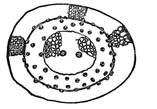
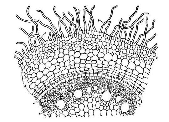
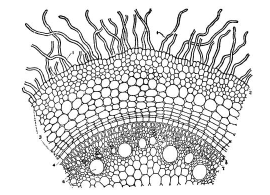
To learn the structure of the roots of grasses we may select as types the roots of Pennisetum cenchroides and Andropogon Sorghum and consider their structural details. In the transverse sections of these roots we find a fairly broad cortex consisting of thin-walled parenchymatous cells more or less regularly arranged. (See figs. 44 and 45.) Just below the piliferous layer two or three layers of thick-walled cells are seen. In the roots of Andropogon Sorghum these thick-walled cells are very conspicuous as they consist of several layers. These layers of thick-walled cells constitute the exodermis. The innermost layer of cells of the cortex is called the endodermis and it becomes conspicuous on account of the thickening in the lateral and inner walls of the cells of this layer.
The rest of the root forming the central core is the stele and at its periphery there is a single layer of cells called the pericycle. The arrangement of the xylem and the phloëm is different from that of the stem. They lie side by side on different radii, and not one behind the other on the same radius as in the stem. The number of xylem groups is fairly large and the development of the xylem is from the pericycle towards the centre of the stele. (See figs. 44 and 45.) The parenchymatous cells in the centre of the stele become thick-walled in older roots.
Structure of the leaf
Structure of the leaf.—The structure of the leaf of grasses is quite characteristic of the family. In every leaf a number of [Pg 33]vascular bundles, some small and others large, pass from the base to the apex. Externally the leaf is covered on both the sides by the epidermis. The spaces existing between the vascular bundles and the epidermis are filled with parenchymatous cells.
The larger vascular bundles consist of xylem and phloëm surrounded by a bundle sheath of a single layer of cells. In the smaller bundles the xylem is very much reduced. Around every vascular bundle there is a single row of somewhat large cells densely packed with large chloroplasts, the chlorophyllous layer. The vascular bundles are strengthened by fibres, on both the sides in the case of larger bundles and on only one side in small bundles.
For a detailed study of the structure of the leaves of grasses the leaf of the grass Panicum javanicum may be chosen. In a transverse section of this leaf, the vascular bundles are very conspicuous. The larger bundles are normal in every way, while in the smaller ones the xylem elements are considerably reduced. Around every one of the vascular bundles there is a single row of large cells containing large chlorophyll grains (the chlorophyllous layer). In a well developed large vascular bundle the chlorophyllous layer is open below just close to the sclerenchymatous band.
On both sides of the larger vascular bundle there are bands of sclerenchyma. In the case of smaller bundles some are strengthened by sclerenchyma on the lower side and others have none. The spaces between the bundles are occupied by thin-walled parenchymatous cells containing small chlorophyll grains.
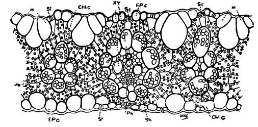
The lower epidermis of the leaf in the transverse section is even and consists of small and large round cells. The upper epidermis is slightly wavy and it is made up of some small round cells alternating with groups of larger cells. The epidermal cells lying over sclerenchyma and the smaller vascular bundles are small and round, while those lying over the furrows between the vascular bundles are large and are called motor or bulliform cells. The presence of motor cells is a characteristic feature of the leaves of many grasses.
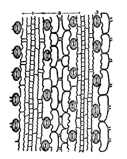
The continuity of both the upper and the lower epidermis is interrupted by the stomata. Air-cavities are seen below these stomata. The arrangement of the stomata, the shape of the guard cells and the characteristics of the epidermal cells become clear on examining a piece of epidermis.
The structure of the leaf of Panicum javanicum may be taken as typical of the structure of the leaves of most grasses. The leaves of Eriochloa polystachya, Cynodon and Paspalums are very much like the leaves of Panicum javanicum in their internal structure. Considerable amount of variation, however, occurs in the leaves of grasses especially as regards the arrangement of fibres and motor cells.
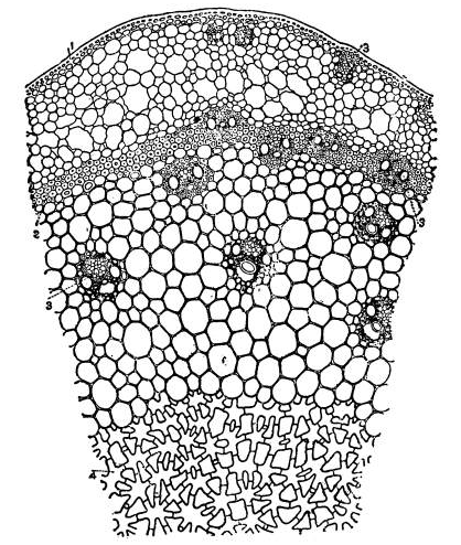
A portion of the transverse section of the leaf of Eriochloa polystachya × 120 1. Motor cell; 2. stomata; 3. sclerenchyma; 4. chlorophyllous layer. Every large primary vascular bundle in the leaves of many grasses possesses sclerenchymatous bands both above and below. The other vascular bundles may have bands of sclerenchyma on both sides or on one side only or none. For example, in the leaves of Panicum repens both the primary and secondary bundles are provided with sclerenchyma on both the sides, while those of the third order may have it on one side or not. The hyaline margin of this leaf and of the leaves of other grasses consists entirely of sclerenchyma.
All the vascular bundles in the leaves of Aristida setacea have broad sclerenchymatous bands on both the sides. Besides these bands arranged like a girder above and below each bundle, there are on the lower side bands of sclerenchyma. So the sclerenchyma becomes almost continuous on the lower side.
The sclerenchyma lying on the lower side of the primary bundles are contiguous with the bundle, while those above are separated from the bundle by the chlorophyllous layer.In the case of secondary and tertiary bundles the sclerenchymatous [Pg 37]bands lying on the lower side are in contact with the chlorophyllous layer, whereas the upper bands are either in contact with this layer or separated from it by a few parenchymatous cells.
All the vascular bundles in the leaves of Eragrostis Willdenoviana are provided with sclerenchyma on both the sides.
The lower band of the primary vascular bundles is continuous with the vascular bundle, the chlorophyllous layer being open below. The upper bands of the primary and the lower bands of the secondary vascular bundles just touch the chlorophyllous layer. In the secondary bundles the sclerenchyma band above is separated from the chlorophyllous layer by two layers of parenchyma. In the case of the leaves of Panicum flavidum, P. colonum, P. fluitans and Pennisetum cenchroides the sclerenchyma is separated from the chlorophyllous layer by layers of parenchyma.
Even from the few examples dealt with above, it is obvious that the range of variation of sclerenchyma in leaves is very great. In the leaves of Aristida setacea there is a considerable amount of sclerenchyma whilst in some leaves such as those of Panicum[Pg 38] colonum, P. flavidum and Panicum fluitans the sclerenchyma is reduced to its minimum.
In the leaves of grasses growing in dry situations the development of sclerenchyma is generally very considerable. The grass Aristida setacea is a good example of a xerophytic grass. The sea-shore grass Spinifex squarrosus is another example of the same kind. But in the leaves of this grass, the development of sclerenchyma is not very considerable, but there is a great development of parenchymatous cells free from chlorophyll within the leaf, the chlorophyll bearing cells being confined to the upper and the lower surfaces of the leaves.
The upper and the lower surfaces of the leaves of many grasses are more or less even, but in the case of a few grasses the upper surface consists of ridges and furrows, instead of being even. In the leaves of Panicum repens and Eragrostis Willdenoviana the upper surface is wavy and consists of shallow furrows and slightly raised ridges. But in the leaves of Aristida setacea and Panicum fluitans the furrows are deeper and the ridges are more prominent. In Aristida setacea the ridges are flat-topped and they are rounded with broad furrows in Panicum fluitans.
The epidermis covering the leaves consists of elongated cells with plane or sinuous walls, various kinds of short cells intercalated between the ends of long cells, motor-cells and stomata. Hairs of different sorts occur as outgrowths of the epidermis. The roughness of the surface of the leaves of grasses is due to the presence of very minute short hairs borne by the epidermis. In most cases these short hairs are found in regular rows. Although the epidermis is more or less even in the leaves of several grasses such as Panicum repens, P. flavidum and Eriochloa polystachya, it is wavy or undulating in the leaves of a few grasses. For example, the upper epidermis in the leaves of Panicum fluitans is undulating as it follows the contour of the ridges and furrows.
The epidermal cells have even surfaces in the leaves of most grasses but in some they bulge out. In the leaves of Panicum flavidum the cells of the lower epidermis are quite even, whilst those of the upper epidermis bulge out. The cells of both the upper and the lower epidermis are distinctly bulging out in the [Pg 40]leaves of Panicum colonum. In Panicum fluitans the cells of the upper epidermis bulge out so much as to form distinct papillæ.
The free surface of the epidermis is more or less cutinised in the leaves of all grasses. In some leaves the cuticle is very thick and even papillate as in the leaves of Aristida setacea and Panicum repens whilst in others it is very thin, as in the leaves of Panicum colonum and P. fluitans. Cutinisation is rather prominent in the leaves of grasses growing under dry conditions and it is less pronounced in mesophytic grasses.
As regards size, the epidermal cells overlying the sclerenchyma are small and those lying over parenchyma are larger. Amongst the larger cells some may be motor-cells. The stomata occur in regular rows between the vascular bundles and they are quite characteristic of grasses. They are more or less similar in structure in all grasses. In the leaves of many grasses stomata are found in both the upper and the lower epidermis and they are confined to the lower epidermis in a few grasses only.
The motor-cells vary very much both as regards their shape and position. In some leaves as in the leaves of the grass Panicum flavidum the motor-cells are confined to the midrib on the upper surface.
The epidermal cells of this leaf are large and uniformly round.
In the case of most grasses the motor-cells are found in groups of three, four or five between the vascular bundles.
The central motor-cell is usually the largest and it is somewhat obovate in shape in a transverse section of the leaf. In the leaves of Panicum javanicum and Eriochloa polystachya there are three or four motor cells in the group and the group consists of four, five or rarely six motor cells in the leaves of Eragrostis Willdenoviana. When there are distinct furrows between ridges these cells lie in the furrows and they are many in number. In the leaves of Panicum repens there are five to seven motor-cells in the furrows and the single row of cells stretched between the motor-cells and the lower epidermis in the furrow consists of more or less clear cells with sparsely scattered small chlorophyll grains.
The motor-cells occupying the furrows in the leaves of Aristida setacea are more in number than in Panicum repens and are of a different shape. All the cells lying in the furrow between the motor-cells and the sclerenchyma are clear cells free from chlorophyll grains.
Although the motor-cells differ in shape from the ordinary epidermal cells in most grasses, there are, however, a few grasses in which the motor-cells do not differ very much from the epidermal cells except in size. For example, in the leaves of Panicum colonum the motor-cells are just like the ordinary epidermal cells in shape but are larger.
Motor-cells are usually confined to the upper epidermis, but they may also be found in the lower epidermis. In the leaves of Pennisetum cenchroides motor-cells are found in both the upper and the lower epidermis, the group in the upper epidermis alternating with that in the lower.
